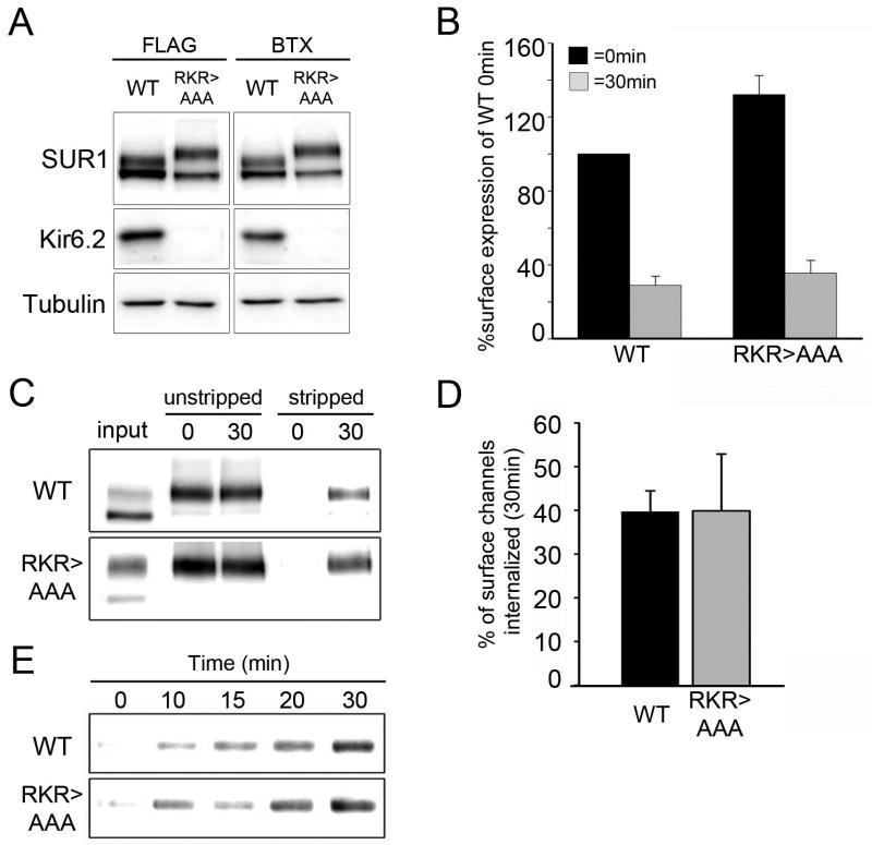
Figure 4. Biochemical analysis of SUR1RKR→AAA internalization.
(A) Western Blot of WT FLAG- or BTX tag-SUR1/ Kir6.2 and FLAG- or BTX-SUR1RKR→AAA expressed alone. Protein expression levels of WT and SUR1RKR→AAA are comparable whether SUR1 is fused with a FLAG-epitope or BTX tag. (B) Monitoring endocytosis of WT and SUR1RKR→AAA by quantitative chemiluminescence assays. Cells expressing WT fSUR1/ Kir6.2 or fSUR1RKR→AAA channels were labeled with anti-FLAG antibody at 4°C followed by a 30min chase at 37°C. FLAG-tagged channels remaining at the cell surface at 30min were detected by chemiluminescence assay and normalized to WT levels at 0min. Internalization was comparable for WT channels and SUR1RKR→AAA (% surface expression for WT: 100 at 0min and 29.0±5.0 at 30min; % surface expression for SUR1RKR→AAA: 132±10.5 at 0min and 35.6±6.9 at 30min; n=4). (C) Monitoring endocytosis of WT and SUR1RKR→AAA by surface biotinylation as described in Materials and Methods. Internalization of WT and SUR1RKR→AAA channels was similar over a 30min time interval when normalized to the total surface label present at 0min (compare 30min stripped samples to 0min unstripped samples). (D) Quantification of WT and SUR1RKR→AAA biotinylation signals by densitometry (n=4). The percentage of surface label internalized at 30min was not significantly different between WT channels (39.7±4.8%) and SUR1RKR→AAA (39.9±12.9%). (E) Time course of internalization assessed by surface biotinylation pulse-chase experiments (n=2). Similar kinetics of internalization was observed between WT channels and SUR1RKR→AAA.

