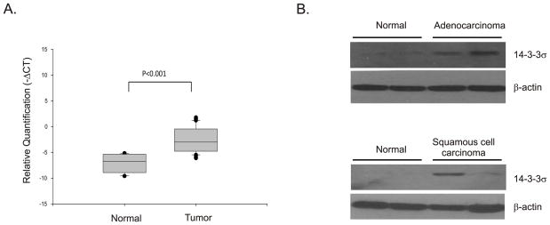
Figure 1. Expression of 14-3-3σ is increased in human lung tumors.
(A) Total RNA was extracted from frozen specimens of normal and tumor tissues and the quantity of 14-3-3σ was determined using quantitative RT-PCR. Relative quantification (−ΔCT) values were determined by comparison with GAPDH. The box plots depict relative quantities of 14-3-3σ in normal and tumor tissues. p-values are for comparison of the levels of 14-3-3σ mRNA in tumor specimens with normal tissue and is calculated using ΔCt values. (B) Protein extracts prepared from the same tumor samples as in A were fractionated on SDS-PAGE protein gels, blotted and probed for 14-3-3σ protein using mouse monoclonal 14-3-3σ antibody (sc-100638, Santa Cruz, CA). Typical results for four normal and four tumor specimens are shown. β-actin was used as a loading control.

