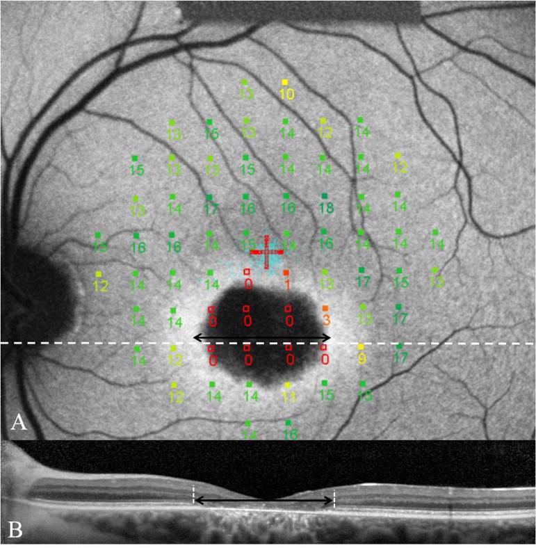
FIG. 6.
Image A demonstrates MP-1 results of a 68 point 10-2 field superimposed and registered to an FAF image of a patient with BEM secondary to mutations in ABCA4. This patient has superior eccentric fixation with the PRL located away from the hyperfluorescent transition zone. Note the profound reduction in sensitivities overlying the atrophic area as well as the reduced sensitivity over the transition zone. The SD-OCT image corresponds to the white dashed line. The black double headed arrow indicates the extent of loss of ISOS junction on the SD-OCT image and is duplicated on the AF image. This demonstrates loss of ISOS junction extending into regions overlying structurally preserved RPE. 152×153mm (300 × 300 DPI)

