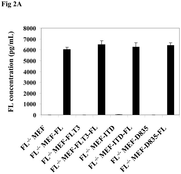
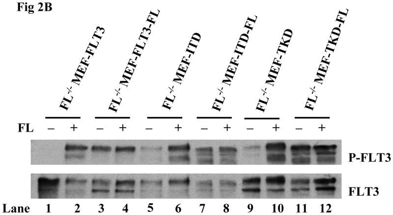
Fig 2. Stable FL expression results in enhanced FLT3 phosphorylation in FL−/− MEF cells expressing FLT3/ITD and FLT3/TKD mutants.
(A) FL−/− MEF cells expressing wt and mutant forms of FLT3 with and without stable expression of FL were washed with PBS, plated at 5×10⁵ cells/mL, and cultured for 24 hours in serum-free medium. Cell culture supernatant was then collected by centrifugation and FL concentration was assessed by ELISA assay.
(B) Cells were cultured in fresh serum free medium for 16 hours and washed with PBS once before stimulation with FL (100 ng/mL for 15 minutes). Total cellular protein extracts derived from 1 × 10⁷ cells were immunoprecipitated with anti-FLT3 antibody. The immunoprecipitates were resolved by 8% SDS-PAGE and subjected to immunoblot analysis with the antiphosphotyrosine antibody 4G10 (top panel). The same blot was stripped and reprobed with anti-FLT3 antibody (bottom panel).


