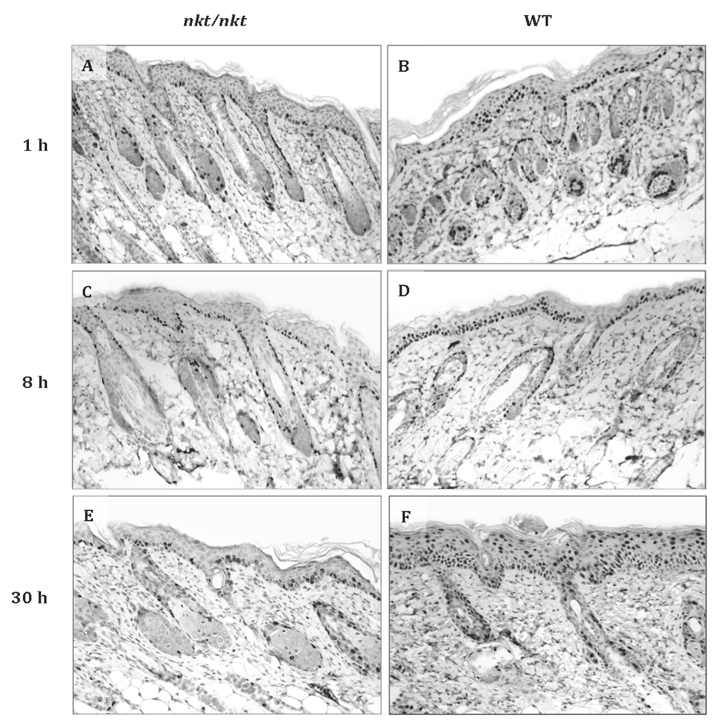
Figure 5. Keratinocyte transit after TPA treatment.

Representative BrdU immunostaining of TPA-treated dorsal skin from a DBA/2-nkt/nkt mouse and a wild-type littermate after 1 h (A and B), 8 h (C and D), and 30 h (E and F) of BrdU injection (all magnifications ×100). Mice were injected i.p. with BrdU 17 h after the last of four TPA applications (3.4 nmol) over a two week period and sacrificed 1, 8, and 30 h after BrdU injection. A total of 4 mice per genotype per time point were evaluated. At 8 h and 30 h after BrdU injection, progression of BrdU-labeled keratinocytes towards the surface is clearly slower in the mutant epidermis. Percentages of BrdU-labeled nuclei in the basal and suprabasal layer for the three time points are shown in Table 1.
