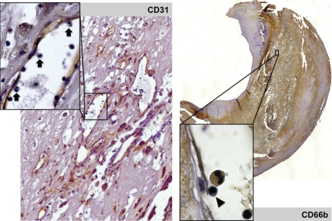
Figure 5.
(Left panel) Immunostaining of endothelium (CD 31, ×20) showing the abundance of neovessels, and the presence of rolling polymorphonuclear cells within these neo-venules (inset, ×40, arrows). (Right panel) Diffuse immunostaining of neutrophil membranes (CD 66b, mosaic reconstituted section, ×2.5) and presence of a rolling CD 66+ polymorphonuclear cell and a mononucleate cell (arrow head) in a neovessel (inset, ×100).

