
Figure 8.
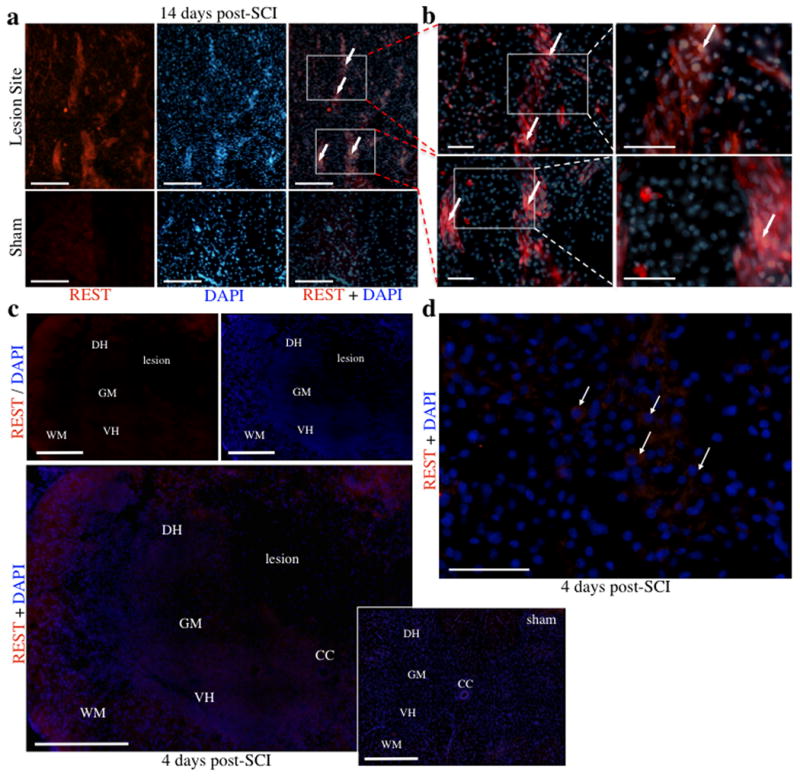
Expression of REST is increased following SCI. a, REST expression at 14 days was increased in cells within and adjacent to the lesion site, but not in sham controls. b, Boxed in sections from (a) are shown in higher magnification in adjacent panels (20× and 40×, respectively). c, Expression of REST at 4 days increased within both white and grey matter of coronal sections, including the dorsal and ventral horns. d, High magnification micrograph shows cytoplasmic REST expression in large neuronal cells (indicated by arrows) of a contused specimen 4 days post-SCI. Abbreviations, central canal (CC), dorsal horn (DH), grey matter (GM), ventral horn (VH), and white matter (WH). Scale bars, 500 μm (c), 200 μm (a) and 50 μm (b, d).