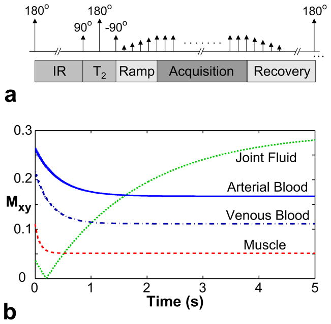
Figure 4.
a: The pulse sequence diagram of a magnetization-prepared bSSFP sequence. The preparation section, which consists of inversion recovery, T2-preparation, and catalyzation segments, is followed by bSSFP acquisitions. Afterwards, the magnetization is allowed to recover, and the whole module is repeated to acquire the next k-space segment. b: The magnetization-prepared transient signals (i.e., during the acquisition period) for arterial blood (T1/T2 =1273/254 ms), venous blood (1273/159 ms), muscle (870/50 ms), and joint fluid (2850/1210 ms).

