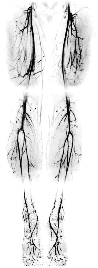
Figure 7.
A healthy 28-year-old female subject was examined to produce IDEAL bSSFP angiograms of the entire lower extremities in three stations: the thigh, calf, and foot respectively. Detailed depiction of the peripheral vasculature is achieved through high-resolution acquisitions and reliable background suppression. The ‘gray’ background in the images is due to the residual signal from peripheral muscle tissue, which is closer to the coils than the deep arteries. This creates an inadvertent increase in the muscle signal.

