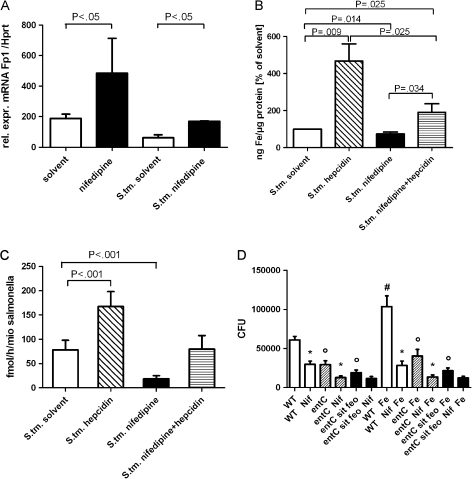
Figure 2.
Effects of nifedipine (Nif) on iron gene regulation, intracellular iron content, and iron acquisition in Salmonella enterica serovar Typhimurium (S. tm.) infection. Experiments were performed as described in the text. Fpn1 messenger RNA (mRNA) levels of macrophages were determined by quantitative reverse-transcription polymerase chain reaction. Values were normalized to the levels of housekeeping gene hypoxanthine phosphoribosyl transferase (Hprt) mRNA (A). The intracellular iron content of infected and treated RAW264.7 cells was determined by means of atomic absorption spectrometry. Iron content was normalized to micrograms of protein of cell pellets (B ). Iron uptake by S. Typhimurium was determined by measuring 59Fe incorporation of bacteria in fmol per hour per one million (mio) Salmonella (C ). RAW264.7 cells were infected with different strains of S. Typhimurium: wild type (WT) (white bars), entC single mutant (shaded bars), and entC sit feo triple mutant (black bars). Differences between the entC or entC sit feo strains and the WT solvent control with or without iron are indicated by circles (P < .001). Differences in the number of colony-forming units (CFUs) following nifedipine treatment of macrophages infected with WT and entC mutant bacteria were compared with respective solvent controls, as indicated by asterisks (P < .001). The difference in the number of CFUs upon iron sulphate addition is indicated by a hash mark (P < .001) (D). Bars represent means (± SD) of 3–6 independent experiments. Calculations for statistical differences between various groups were performed by means of the Student’s t test, Mann--Whitney U test, or multivariate analysis with the Bonferroni correction where appropriate. Rel. expr., relative expression.

