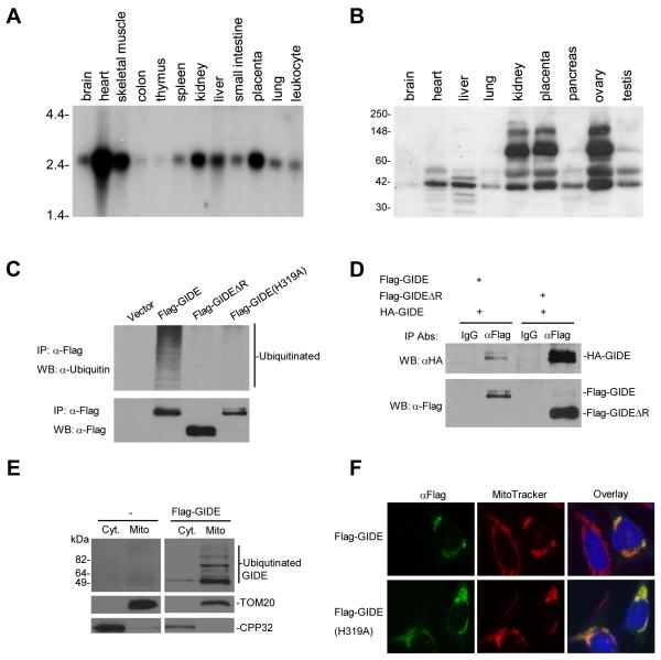
Fig. 2.
Expression, ubiquitination and localization of GIDE. A. Northern blot analysis of human GIDE mRNA expression in various tissues. B. Western blot analysis of human GIDE protein expression in various tissues. C. GIDE is auto-ubiquitinated. 293 cells were transfected with the indicated Flag-tagged plasmids. Cell lysates were immunoprecipitated with anti-Flag antibody, the immunoprecipitates were analyzed by Western blot with anti-ubiquitin (upper panel) and anti-Flag (lower panel) antibodies. D. GIDE forms oligomers. 293 cells were transfected with the indicated GIDE or its mutant plasmids. Cell lysates were immunoprecipitated with anti-Flag antibody or control mouse IgG, the immunoprecipitates were analyzed by Western blot with anti-HA antibody. E. Biochemical study of GIDE localization. 293 cells were transfected with Flag-GIDE or left untransfected for 12 hours. Mitochondrial and cytosolic fractions were isolated and analyzed by Western blots with the indicated antibodies. F. Immunofluorescent staining of GIDE and its mutant GIDE(H319A). HeLa cells were transfected with the indicated plasmids and stained with the indicated antibodies.

