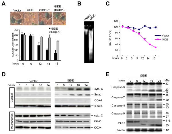
Fig. 3.
GIDE induces apoptosis in 293 cells. A. The effects of GIDE and its mutants on cell survival. 293 cells (~1×10⁵) were transfected with 0.1 μg of CMV-β-gal plasmid and 0.5 μg of the indicated expression plasmids. Sixteen hours after transfection, cells were stained with X-gal and photographed (upper panels), or survived blue cells were counted (lower panel). B. Overexpression of GIDE causes DNA fragmentation. 293 cells (~1×10⁶) were transfected with 10 μg of the indicated plasmids. Sixteen hours after transfection, DNA was isolated from the transfected cells and analyzed by 2% agarose gel. C. Overexpression causes loss of mitochondrial membrane potential. 293 cells (~2×10⁵) were transfected with 1 μg of the indicated expression plasmids. Sixteen hours after transfection, cells were stained with Rh123 (10 μg/ml) at 37 0C for 30 min and then washed with PBS (+) for three times. The mitochondrial membrane potential was analyzed by flow cytometry. D. Overexpression of GIDE causes release of cytochrome C and Smac from mitochondria to cytosol. 293 cells were transfected with GIDE or control plasmid for the indicated times. The cell lysates were fractionated and analyzed by Western blots with the indicated antibodies. E. Overexpression of GIDE causes activation of caspases. 293 cells were transfected with GIDE for the indicated times. Cell lysates were analyzed by Western blots with the indicated antibodies.

