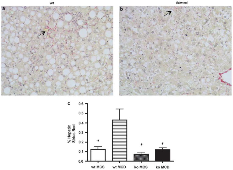
Figure 5.
Gclm null mice are resistant to MCD diet-induced fibrosis. Sirius red-stained section of MCD-fed liver of wt mouse (a) shows pericellular Sirius red staining (arrow), indicating collagen deposition; little staining is detected in MCD-fed Gclm (ko) null liver (b). Morphometric analysis of hepatic Sirius red staining (c). Staining was significantly increased in MCD-fed wt liver (wt MCD) compared with MCS-fed wt mice (wt MCS), and MCS or MCD-fed Gclm null mice (ko MCS; ko MCD). *P <0.05 compared with wt MCD, for all groups, n = 4–6 animals.

