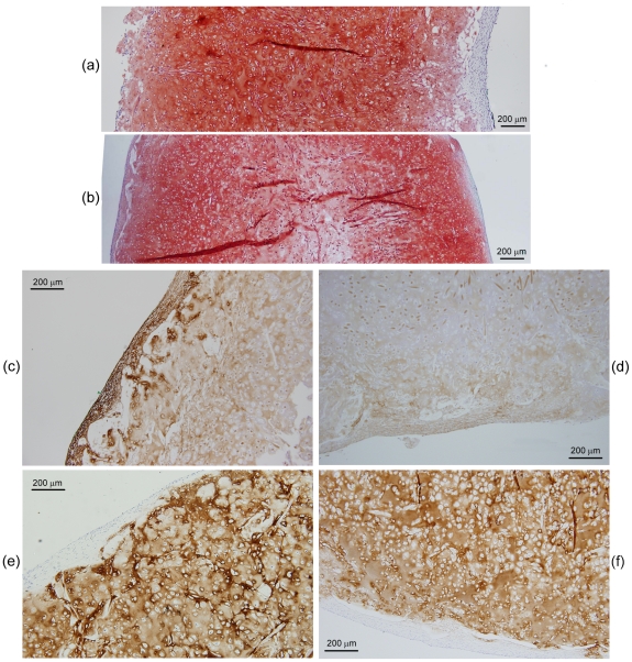
Figure 7. Histological appearance of constructs produced using PGA–alginate scaffolds cultured in bioreactors for 5 weeks at a constant high flow rate of 0.2 mL min−1 (a, c, e) or a gradually increasing flow rate of 0.075–0.2 mL min−1 (b, d, f). The scaffolds were seeded using 20×106 cells.
Construct cross-sections show: (a, b) pink–red staining for GAG, blue staining for collagen, and dark blue–purple staining for cells; (c, d) immunostaining (brown) for collagen type I; and (e, f) immunostaining (brown) for collagen type II.

