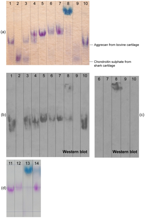
Figure 9. Analysis of proteoglycan size and integrity: (a and d) results from electrophoresis on composite acrylamide–agarose gels; (b and c) results from Western blots probed using monoclonal antibody specific to the hyaluronic-acid-binding region of human aggrecan.
The back-up nitrocellulose membrane for capture of smaller-sized molecules is shown in (b); the primary membrane showing larger-sized molecules is shown in (c). Lane 1 – aggrecan from bovine cartilage; Lane 2 – chondroitin sulphate from shark cartilage; Lane 3 – a 1∶1 w/w mixture of bovine aggrecan and chondroitin sulphate; Lane 4 – a 1∶1 w/w mixture of bovine aggrecan and proteoglycans isolated from human fetal cartilage; Lane 5 – proteoglycans isolated from human fetal cartilage; Lanes 6 and 7 –proteoglycans isolated from tissue-engineered cartilage; Lane 8 – spent medium from bioreactor culture of tissue-engineered cartilage; Lane 9 – bovine aggrecan digested with papain; Lane 10 – aggrecan from bovine cartilage; Lane 11 – proteoglycans isolated from human fetal cartilage; Lanes 12, 13 and 14 – spent medium from bioreactor culture of tissue-engineered cartilage. The sample in Lane 12 was diluted and treated with 6 M urea; the sample in Lane 14 was treated with hyaluronidase.

