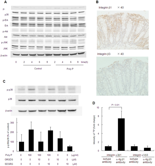
Figure 7. Poly P interacts with integrin β1 and activates p38 MAPK pathway.
(A) Western blot analysis of p38, ERK, JNK and Akt phosphorylation in Caco2/BBE by 10 µg/mL of poly P. (B) Expression and localization of integrin β1 and β3 in mouse small intestine. Immunohistochemistry for integrin β1 and β3 were performed by using anti-integrin β1, rabbit-poly antibody and anti-integrin β3, rabbit-poly antibody. (C) Inhibition of poly P-induced p38 MAPK phosphorylation by RGD peptide. C57Bl/6 mice small intestinal loops were filled with RPMI 1640 medium containing 10 µg/mL of poly P, 10 µM of Gly-Arg-Gly-Asp-Ser (GRGDS) or Ser-Asp-Gly-Arg-Gly (SDGRG) 2 h at 37°C in a 5% CO2 incubator. Their p38 MAPK phosphorylation was detected by western blot (n = 3, each group). (D) Poly P-integrin β1 or β3 interaction analysis. Reaction mixture of 32P-labeled poly P and integrin α3β1 or αVβ3 was immunoprecipitated with integrin β1 or β3 antibody and protein G beads. Immunoprecipitated 32P-labeled poly P was determined with liquid scintillation counting.

