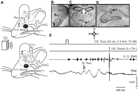
Figure 1. Experimental design.
(A) EMG recording electrodes were implanted in the orbicularis oculi (O.O.) muscle of the upper left eyelid. In addition, bipolar stimulating electrodes were implanted on the ipsilateral supraorbital nerve for presentation of unconditioned stimulus (US). The conditioned stimulus (CS) consisted of a tone delivered from a loudspeaker located 30 cm from the animal's head. Animals were also implanted with stimulating electrodes in the thalamic reuniens nucleus and with recording electrodes in the medial prefrontal cortex (top diagram) or the hippocampal CA1 area (bottom diagram). (B–D) Photomicrographs illustrating the location of recording electrodes in the mPFC (B) and in the hippocampal CA1 area (D), as well as the stimulation (C) site (arrows). Calibration bars 500 µm. Abbreviations: D, L, M, V, dorsal, lateral, medial, and ventral. E, A schematic representation of the conditioning paradigm, illustrating CS and US stimuli, and the moment at which a single pulse (100 µs, square, biphasic) was presented to the reuniens nucleus (St. Reu.). An example of an EMG record from the orbicularis oculi (O.O.) muscle obtained from the 9th conditioning session is illustrated, as well as an extracellular record of hippocampal activity from the same animal, session, and trial. Note the fEPSPs evoked by the pulse presented to the reuniens nucleus.

