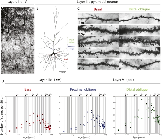
Fig. 1.
(A) Representative low-magnification photographs of the rapid Golgi-impregnated layer IIIc and V pyramidal cells in the dorsolateral prefrontal cortex of a 16-y-old subject. Black arrows indicate basal dendrites, and gray arrows indicate oblique dendrites. (Scale bar: 100 μm.) (B) Neurolucida reconstruction of layer IIIc pyramidal neuron of a 49-y-old subject, illustrating sites selected for counting spines over a 50-μm length of apical distal oblique dendrites (green), apical proximal oblique dendrites (blue), and basal dendrites (red). (Scale bar: 100 μm.) (C) Representative high-power magnification images of rapid Golgi-impregnated layer IIIc pyramidal neurons from the dorsolateral prefrontal cortex showing basal dendrites (Left) and distal apical oblique dendrites (Right) during different stages: an infant 1 mo of age, a 2.5-y-old child, and 16-y-old, 28-y-old, and 49-y-old subjects. (Scale bar: 10 μm.) (D) Graphs representing number of dendritic spines per 50-μm dendrite segment on basal dendrites after the first bifurcation (red); apical proximal oblique dendrites originating within 100 μm from the apical main shaft (blue); and apical distal oblique dendrites originating within the second 100-μm segment from the apical main shaft (green) of layer IIIc (filled symbols) and layer V (open symbols) pyramidal cells in the dorsolateral prefrontal cortex. Squares represent males; circles represent females. The age in postnatal years is shown on a logarithmic scale. Puberty is marked by a shaded bar. B, birth (fourth postnatal day); P, puberty. Specification of tissue analyzed is given in Table S1, and the positions of sections on which pyramidal neurons were measured are indicated on the reconstructed pyramidal neuron shown in B.

