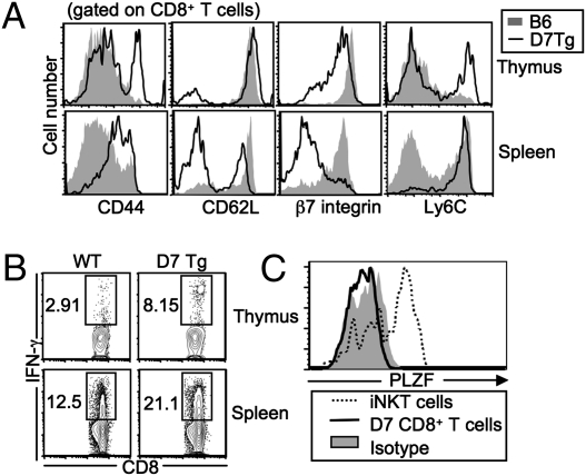
Fig. 1.
Characterization of CD8+ T cells in D7 Tg mice. (A) Splenocytes and thymocytes from D7+Rag−/− and B6 mice were stained with mAb against various lymphocyte activation markers. Data shown are representative of three to six mice in each group. (B) Ex vivo stimulation of CD8+ T cells from D7+Rag−/− and B6 mice. CD8+ T cells were stimulated with PMA and ionomycin and then subjected to intracellular IFN-γ staining. Data shown are representative of two independent experiments with two mice in each group. (C) PLZF expression on D7 thymocytes, iNKT cells (TCRβ+CD1d/α-galactosylceramide tetramer+), and conventional CD8+ T cells from B6 mice. The expression of PLZF on conventional CD8+ T cells was indistinguishable from isotype control. Data shown are representative of two independent experiments.

