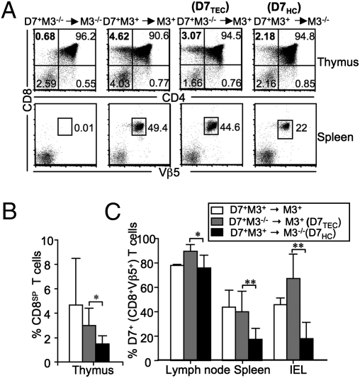
Fig. 2.
Selection of M3-restricted CD8+ T cells on HCs and TECs. Thymocytes and splenocytes from indicated chimeric mice were stained with antibodies specific for CD8β, CD4, and Vβ5. (A) (Upper) Numbers represent percentages of total thymocyte population within each quadrant. (Lower) Percentages of CD8+Vβ5+ cells within the total splenocyte population. Because both donor and recipient mice are on the Rag−/− background, all Vβ5+ cells in chimeric mice are D7 T cells. (B) Percentage of CD8 single positive cells within the total thymocyte population from various BM chimeras. (C) Mean percentage of CD8+Vβ5+ cells in the total lymphocyte populations from the lymph node, spleen, and small intestine ± SEM. *P < 0.05; **P < 0.01. Data shown are representative of four independent experiments with two to three mice per group. IEL, intestinal epithelial lymphocytes.

