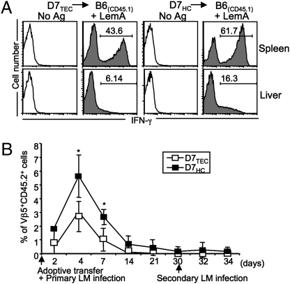
Fig. 4.
D7 T cells selected on TECs and HCs respond with similar kinetics but different magnitudes during LM infection. Sorted D7TECs and D7HCs (CD45.2+) were adoptively transferred into CD45.1 B6 congenic mice and recipient mice infected with LM. (A) Seven days postinfection, splenocytes and hepatic leukocytes from recipient mice were stimulated with LemA. The percentages of cells producing IFN-γ within the CD45.2+CD8+ LemA-specific D7TEC or D7HC population are shown. (B) Percentages of Vβ5+CD45.2+ cells detected in the blood of recipient mice at different time points following primary and secondary LM infection. *P < 0.05. Data shown are representative of two independent experiments with three to four mice per group.

