Fig. 6.
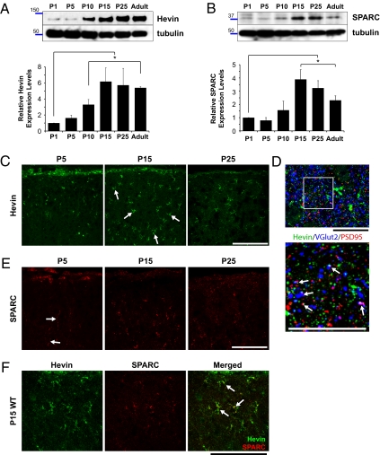
Expression of hevin and SPARC is developmentally regulated at the SC. (A) Upper: Representative Western blot analysis of hevin in SC lysates from P1, P5, P10, P15, P25, and adult mice. Lower: Tubulin as loading control. Graph shows quantification of relative expression levels of hevin protein in SC lysates. The expression levels were quantified first by normalization of the hevin signal to the tubulin signal, and subsequently by calculation of the ratio with respect to mean hevin signal level at P1 (n = 4 animals per age, four separate Western blots; *P < 0.05; error bars indicate SEM; Fig. S6 shows antibody characterization). (B) Upper: Representative Western blot of SPARC protein expression in SC lysates from P1, P5, P10, P15, P25, and adult mice. Lower: Tubulin as loading control. Graph shows quantification of relative expression levels of SPARC protein in SC lysates. The expression levels were quantified first by normalization of the SPARC signal to the tubulin signal, and subsequently by calculation of the ratio with respect to mean SPARC signal level at P1 (n = 4 animals per age, four separate Western blots; *P < 0.05; error bars indicate SEM; Fig. S6 shows antibody characterization). (C) Sagittal mouse brain sections from P5, P15, and P25 mice were stained for hevin with goat anti-hevin polyclonal antibodies (green; R&D Systems). Images are taken from SC. (Scale bars: 100 μm.) (D) High-magnification images from SC that was stained for hevin (12:155; green); a presynaptic marker that is specific for RGC axonal terminals, VGlut2 (blue; Synaptic Systems); and the postsynaptic marker that is specific for excitatory synapses, PSD95 (red; Zymed). (Scale bars: 25 μm.) (E) Sagittal mouse brain sections from P5, P15, and P25 mice were stained for SPARC with goat anti-SPARC polyclonal antibody (red; R&D Systems). Images are taken from SC. (Scale bars: 100 μm.) (F) Hevin and SPARC proteins are colocalized at P15 (SC). Upper: Sagittal P15 mouse brain sections were stained for hevin and SPARC with rat anti-hevin (12:155) and goat anti-SPARC antibodies (R&D Systems). Lower: Hevin and SPARC colocalize in the P15 mouse SC (arrows). (Scale bar: 100 μm.)

