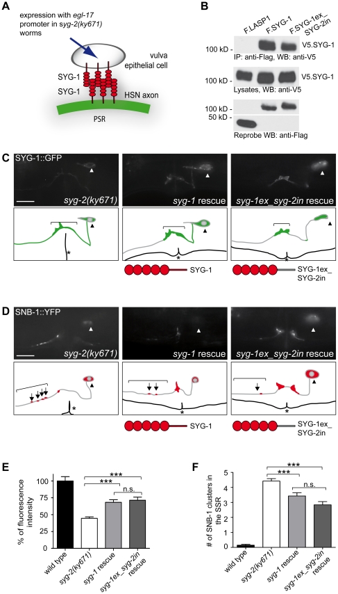
Figure 4. SYG-1 can partially rescue SYG-2 functions in vulva epithelial cells.
A, Schematic representation of SYG-1 transgene expression in the vulva epithelial ecells under the egl-17 promoter. B, SYG-1 interacts with SYG-1 and chimerical SYG-1ex_SYG-2in. V5-tagged SYG-1 and Flag-tagged SYG-1, SYG-1ex_SYG-2in, or LASP1 were co-expressed in HEK 293T cells and precipitated with anti-Flag antibody. V5.SYG-1 bound to SYG-1 and chimerical SYG-1ex_SYG-2in was detected using anti-V5 antibody The control protein LASP-1 failed to immobilize SYG-1 (upper panel). The middle panel shows expression of V5.SYG-1 in the lysates, the lower panel expression of the Flag-tagged proteins. IP, immunoprecipitation. WB, Western blot. kD, kilodalton. C, Ectopically expressed syg-1 and syg-1ex_syg-2in in syg-2(ky671) mutants increases localization of SYG-1::GFP to the PSR in HSN compared to mutant animals. D, Ectopic expression of syg-1 and syg-1ex_syg-2in in vulva epithelial cell of syg-2(ky671) mutants reduces SNB-1 clusters in the SSR. Fluorescence image and schematic drawing. Arrowhead, HSN cell body. Arrow, SNB-1 cluster. Asteriks, vulva. Scale bar represents 10 µm. Lateral view, anterior is to the left, ventral down. E, Quantification of fluorescence intensity of SYG-1::GFP in the PSR. Ectopic expression of syg-1 and syg-1ex_syg-2in partially rescues the syg-2(ky671) mutant phenotype. Student t-test/Mann-Whitney rank sum test. ***, p<0,001. n.s., not significant. n>20. Error bars, SEM. F, Quantification of SNB-1::YFP punctae in the SSR. Ectopic expression of syg-1 or syg-1ex_syg-2in partially reduces the syg-2(ky671) mutant phenotype. Mann-Whitney rank sum test. ***, p<0.001. n.s., not significant. n>50. Error bars, SEM.

