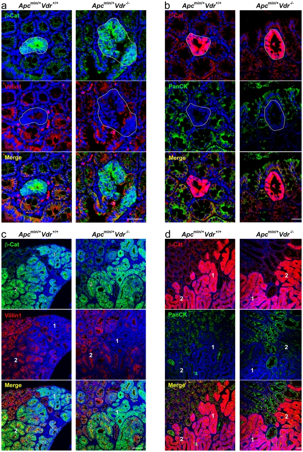
Figure 6. Vdr depletion does not affect the differentiation status of colon ACF and carcinomas in Apcmin/+ mice.
Representative immunofluorescence images showing β-catenin and villin1 (a, c) or β-catenin and pan-cytokeratin (b, d) expression and localization in colon ACF (a, b) and carcinomas (c, d) from Apcmin/+Vdr+/+ and Apcmin/+Vdr-/- mice. Dotted-lines delimit ACF. Numbers indicate regions with different pattern of expression of the analyzed proteins within the same tumor: 1) high β-catenin, low villin1/cytokeratins; 2) low β-catenin, high villin1/cytokeratins. Scale bars, 50 µm (a, b) and 200 µm (c, d).

