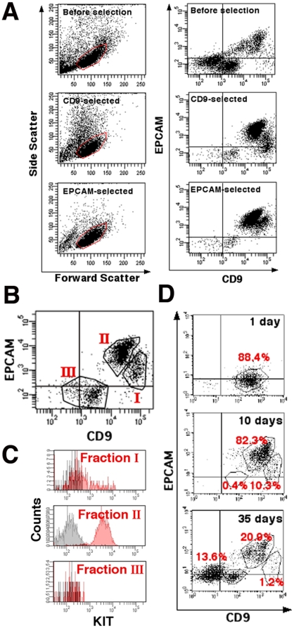
Figure 2. Flow cytometric analyses of CD9- or EPCAM-selected cells after MACS.
(A) Light-scattering properties and double immunostaining of total testis cells (Top), CD9-selected cells (Middle) or EPCAM-selected cells (Bottom) stained with APC-conjugated anti-CD9 and PE-conjugated anti-EPCAM antibodies. Cells were gated according to forward scatter (size) and side scatter (cell complexity) values (Left). Gated cells were analyzed for CD9 and EPCAM (Right). Note the simpler light-scattering properties of EPCAM-selected cells. (B) Three subpopulations of CD9-selected cells. Fraction I shows high CD9 and low or no EPCAM immunostaining; fraction II shows low CD9 and high EPCAM immunostaining; and fraction III is low or no CD9 and no EPCAM immunostaining. (C) The three CD9-selected cell subpopulations, immunostained with APC-conjugated anti-CD9, PE-conjugated anti-EPCAM, and PE-Cy7-conjugated anti-KIT antibodies. KIT is strongly expressed in fraction II. Areas shaded in black indicate control staining. (D) Changes in immunostaining of total testis cells during postnatal testicular development. Total testis cells were stained with APC-conjugated anti-CD9 and PE-conjugated anti-EPCAM antibodies. Stronger CD9 and EPCAM immunostaining is seen in 10-day-old mouse testis cells compared with 1-day-old mouse testis cells. Only testes from 35-day-old mice show all three fractions.

