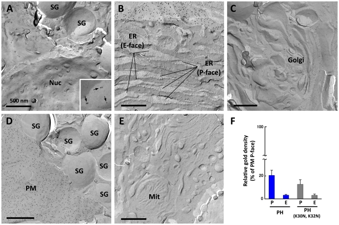
Figure 5. PI(4,5)P2 in intracellular organelles.
(A-E) Organelles were identified by morphological criteria. The nuclear membrane (Nuc) (A), the ER (B), the Golgi apparatus (C), the secretory granule (SG) (A and D), and the mitochondrion (Mit) (E) were devoid of specific labeling for PI(4,5)P2. More colloidal gold particles were seen in the P face of the secretory granule than the other organelles, but they were observed in a similar density even when GST-PH was replaced with GST-PH (K30N, K32N) (Fig. 2B). The inset (A) is an enlargement of a small portion of the P face of the secretory granule membrane to show the presence of non-specific labeling (arrows). (F) Quantification of the relative labeling density (average ± standard error). The labeling density in the secretory granule is shown as the relative ratio to that of the plasma membrane. The data were collected from three independent experiments and areas more than 9.3 µm2 was measured. An equivalent number of colloidal gold particles were observed in the P face of the secretory granule when either GST-PH or GST-PH (K30N, K32N) was used. Therefore, we concluded that the labeling in the secretory granule was insignificant.

