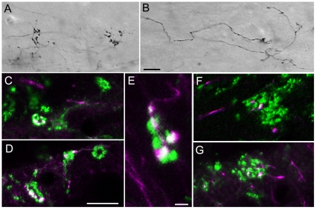
Figure 1. Convergence of tectopulvinar terminals.
Injections of biotinylated dextran amine (BDA) in the SC labels tectopulvinar axons that form widespread (“diffuse”) axons and boutons as well as more discrete clustered (“specific”) boutons. Representative images of BDA-labeled “diffuse” (A) and “specific” (B) axons in 50 µm thick sections are illustrated using transmitted light, a 40x objective, and Nomarski optics. C–G) Confocal images (single 0.1 µm scan with a 100x objective) illustrate tectopulvinar terminals labeled with BDA (purple) and tectopulvinar terminals labeled with antibodies against the type 2 vesicular glutamate transporter (vGLUT2, green). Terminals labeled with both BDA and vGLUT2 appear white. Clusters of vGLUT2-stained terminals contain at most 1 bouton contributed by “diffuse” axons (F), while “specific” axons contributed several boutons to each cluster (C-E, G) Scale in B = 10 µm and applies to A. Scale in D = 10 µm and applies to C, F and G. Scale in E = 2 µm.

