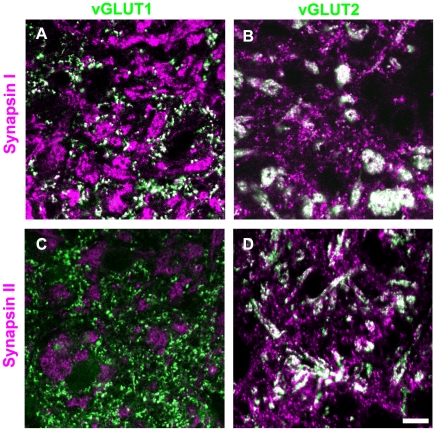
Figure 4. Distribution of synapsins and vGLUTs in the pulvinar nucleus.
Tree shrew pulvinar tissue was labeled with antibodies against synapsin I or II (purple), and vGLUT1 or 2 (green). Profiles that are labeled with two antibodies appear white. Representative confocal images of the Pd (single 0.2 µm scan with a 100x objective) are illustrated. Corticopulvinar terminals (RS profiles labeled with the vGLUT1 antibody) contain synapsin I (A), but not synapsin II (C). Tectopulvinar terminals (large clusters of RM profiles labeled with the vGLUT2 antibody) contain both synapsin I (B) and synapsin II (D). Scale = 10 µm.

