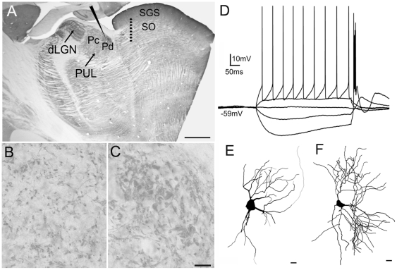
Figure 5. In vitro recording methods.
A) A parasaggital section of the tree shrew brain stained for the type 2 vesicular glutamate transporter (vGLUT2) illustrates the location of the whole cell recordings in the caudal pulvinar nucleus and the location of the 8 electrode stimulus array in the stratum griseum superficial (SGS) and stratum opticum (SO) of the superior colliculus. B, C) High magnification views of the Pc (B) and Pd (C) illustrated in panel A. Immunohistochemical staining for vGLUT2 is a marker for tectopulvinar terminals [9] and reveals the distinct arrangements of tectal terminals in the dorsal (Pd) central (Pc) pulvinar nucleus. The Pd contains dense clusters of tectal terminals and tubular clusters line long lengths of dendrites (C). In contrast, the Pc contains smaller more sparsely distributed clusters of tectal terminals (B). D) Voltage fluctuations recorded in response to the injection of depolarizing or hyperpolarizing current steps of varying size revealed that all recorded cells fired with both tonic action potentials, and low threshold calcium bursts. E) Drawing of a biocytin-labeled cell recorded in the juvenile pulvinar, F) Drawing of a biocytin-labeled cell recorded in the adult pulvinar. Scale in A = 1 mm. Scale in C = 30 µm and also applies to B. Scales in E and F = 10 µm. dLGN, dorsal lateral geniculate nucleus.

