Fig. 3.
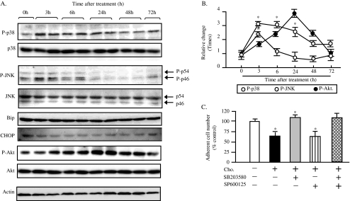
SAPKs are activated by cholesterol loading to MIN6. Whole cell lysates were prepared at 0, 3, 6,24, 48, and 72 h after the exposure to 250 times diluted CLC, and subjected to Western blot analysis using antibodies against phospho-T180/Y182 p38 MAPK (P-p38), total p38 MAPK (p38), phospho-T183/Y185 JNK (P-JNK, P-p54,P-p46), total JNK (JNK, p54,P46), Bip, CHOP, phospho-S347 Akt(P-Akt), total Akt(Akt) and actin. The representative images are shown in a. The quantitative results are shown in b. The phospho-kinase levels were normalized by the actin levels, and are expressed as relative changes of the level at 0 time point. Mean ± SD (n = 3). *, #p < 0.05 vs. the 0 point. The cells were loaded by the cholesterol alone or by the combination of cholesterol and SAPK inhibitors as indicated manner for 24 h. SB203580, 10 μM; SP600125, 10 μM. The number of adherent cells were then counted after removing the detached cells and trypsinization, and expressed as percentage of the number of cholesterol-nonloaded control cells (c). Mean ± SD (n = 3). *p < 0.05 vs. the levels of control

