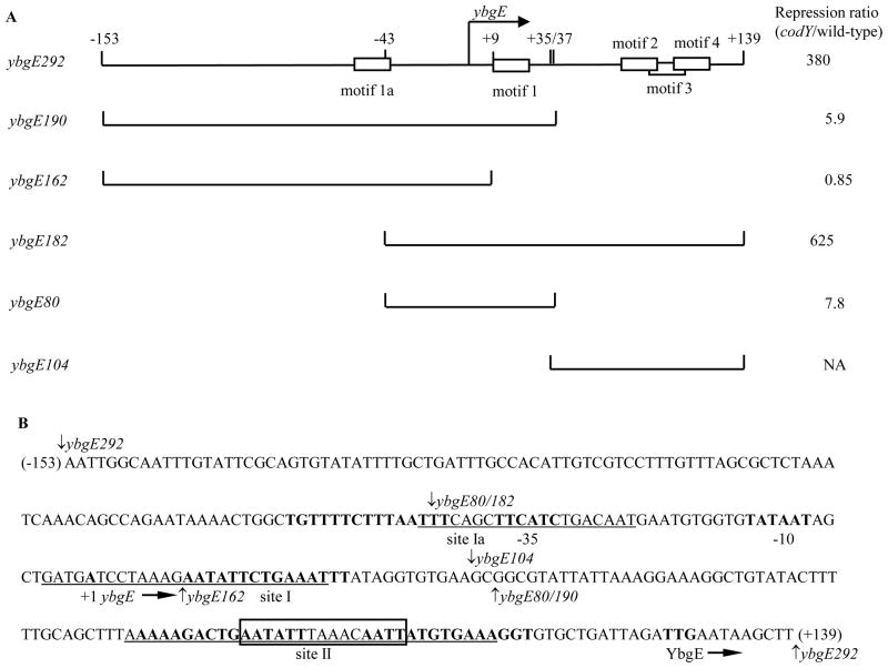
Fig. 1. Plasmid maps and the sequence of the ybgE regulatory region.
A. Schematic maps of the ybgE fragments used in this work. The location of the transcription start point is indicated by the bent arrow. CodY-binding motifs are shown as rectangles. The coordinates indicate the boundaries of different fragments with respect to the transcription start point. The repression ratio is the ratio of expression values for the corresponding lacZ fusions in codY null mutant and wild-type strains in the 16 amino acid-containing medium.
NA – not applicable. The ybgE104 fragment does not contain the promoter and therefore was not tested as part of a fusion.
B. The sequence of the coding (non-template) strand of the ybgE regulatory region. The likely initiation codon, −10 and −35 promoter regions, transcription start site and CodY-binding motifs (15-bp sequences similar to the proposed CodY-binding consensus) 1a, 1, 2, and 4 are in bold. CodY-binding motifs 3 is boxed. The direction of transcription and translation is indicated by the arrows. The sequences protected by CodY (sites Ia, I, and II) in DNase I footprinting experiments on the template strand of DNA are underlined. The boundaries of DNA fragments used in this work are indicated by vertical arrows. The coordinates of the 5′ and 3′ end of the sequence with respect to the transcription start point are shown in parenthesis.

