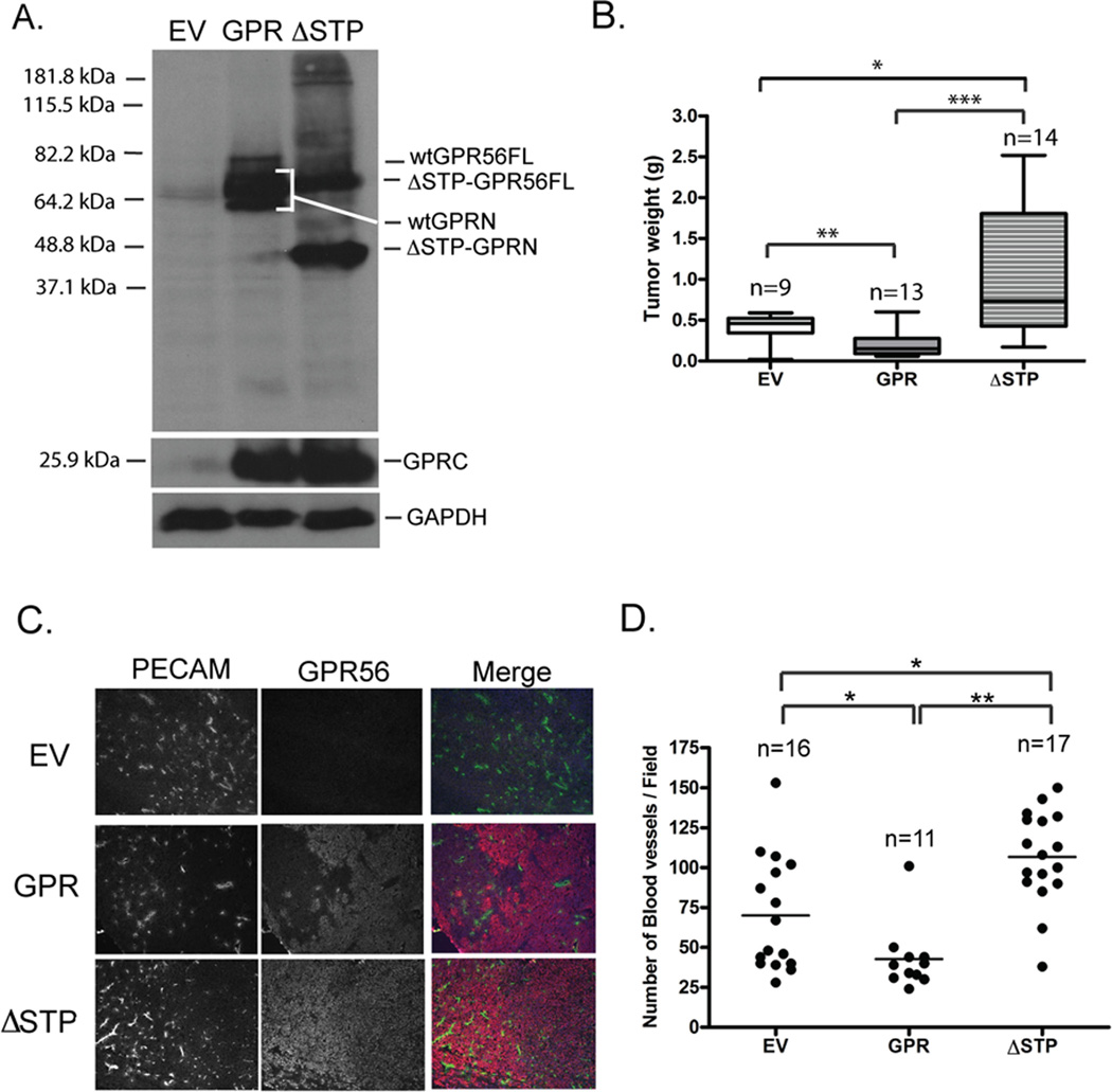
Figure 3. Deletion of the STP segment in GPR56 led to significantly enhanced tumor growth and angiogenesis.
A. Western blot analyses of the expression of ΔSTP-GPR56 in MC-1 cells.
B. Expression of ΔSTP-GPR56 led to enhanced tumor growth. *: p < 0.05; **: p < 0.01; ***: p < 0.001. Student’s t-test.
C. Expression of ΔSTP-GPR56 led to enhanced tumor angiogenesis. Tumor sections were stained with the anti-PECAM antibody (green) and the anti-GPRC antibody (red).
D. Blood vessel density on sections stained with anti-PECAM antibody was quantified as described in the legend for Figure 2. *: p < 0.05; **: p < 0.01; ***: p < 0.001. Student’s t-test.

