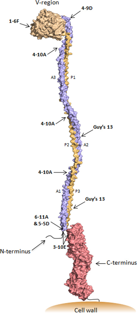
Figure 6.
Schematic representation of the tertiary structure of Antigen I/II. Approximate binding sites of anti-AgI/II (P1) MAbs are indicated. The modeled structure was deduced based on the solved crystal structure of the A3VP1 fragment coupled with the estimated dimensions of the full-length molecule determined by velocity centrifugation [50] and the solved crystal structure of the complete C-terminus [49]. The extended stalk formed by the interaction of the three alanine-rich repeats (blue) with the three proline-rich repeats (orange) brings the pre-A-region into close proximity with the globular C-terminal region. AgI/II family molecules are anchored to the cell wall via sortase following cleavage at the C-terminal LPXTG consensus motif [61].

