
Fig. 1.
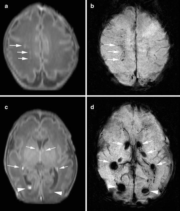
Punctate white matter lesions in a preterm infant born at gestational age of 31 weeks, scanned on day 3 (case 9): GMH-IVH pattern. a T1-weighted image shows punctate hypersignal lesions in the white matter (arrows). b SWI at the corresponding slice to a shows signal loss at the punctate hypersignal lesions on T1-weighted image, suggesting the presence of hemorrhage (arrows). c T1-weighted image at the level of the basal ganglia shows germinal matrix hemorrhage (arrows) and intraventricular hemorrhage (arrowhead) as hypersignal lesions. d SWI at the corresponding slice to c shows bilateral germinal matrix hemorrhage (arrows) and intraventricular hemorrhage (arrowhead)