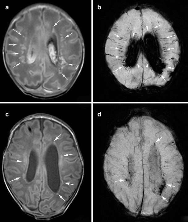
Fig. 3.

Punctate white matter lesions in a preterm infant born at gestational age of 32 weeks born following an emergency cesarean section following a serious car accident of the mother. MRI was performed at 33 and 43 weeks postmenstrual. To manage post-hemorrhagic hydrocephalus a subcutaneous reservoir was placed after the initial scan (case 8). a Initial T1-weighted image shows multiple hypersignal lesions with dotted, nodular and patchy shapes in the white matter (arrows). b Initial SWI at the corresponding slice to a shows signal loss in many of the PWML (arrows). Diffuse signal loss in the lateral ventricles represents large amount of intraventricular hemorrhage (arrowheads). c Follow-up T1-weighted image shows reduced hypersignal in the PWML (arrows). d Follow-up SWI at the corresponding slice to c shows decreased rate of signal loss in PWML (arrows). Note that some of the signal loss at SWI that was visible in the initial scan is no longer seen on the follow-up scan
