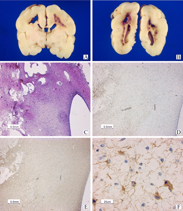
Fig. 5.

Postmortem examination in case 23 (same case in Fig. 4). Macroscopic view of the cerebrum on coronal sections (A, B). Large cystic areas and hemorrhagic changes can be seen in the white matter (C). Low power view of an immunohistological staining using antibodies against CD68 to demonstrate activated microglial cells and macrophages surrounding the cystic and hemorrhagic areas (D). GFAP staining is positive in the periventricular area with abnormalities on T1-weighted imaging, but normal SWI (E low power and F high power of central area in E), indicating early gliosis. No hemorrhages can be demonstrated in this area
