
Figure 4.
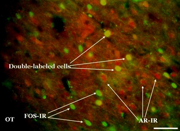
Fos and androgen receptor (AR) double-label sample photomicrograph. Fos and ARs were labeled with monoclonal primary antibodies which were then recognized by fluorescent secondary antibodies. This photomicrograph was taken at the level of the posterior medial amygdala. This image was captured at 20x by a high-resolution black and white camera, and then pseudo-colored (Fos = green, AR = red) using Metamorph software. Scale bar = 100μm.