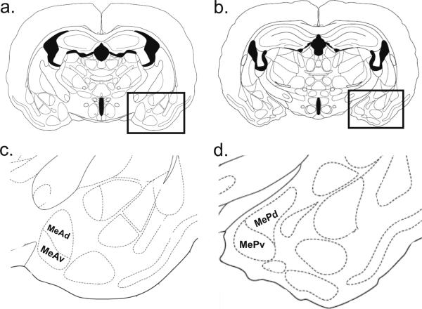
Figure 5.
Schematic of anterior and posterior medial amygdala areas of interest. Coronal sections from the atlas of Morin and Wood (2001) at the level of anterior (a; Bregma: -1.2 mm.) and posterior (b; Bregma: -1.5 mm.) medial amygdala (boxed areas). The borders of the amygdala sub-regions used for counting are displayed in c and d (enlargements of the boxed regions of a and b, respectively).

