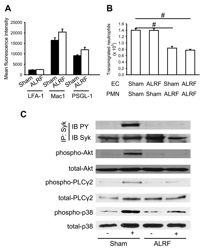
Figure 6. ALRF causes altered intracellular signaling and reduces transmigration in vitro.
(A) Murine neutrophils were isolated from whole blood samples obtained from mice with ALRF and sham operated control mice and FACS analysis was performed to quantify the surface expression of PSGL-1, LFA-1 and Mac-1 (n=3). (B) Isolated neutrophils from mice with ALRF and sham operated control mice were allowed to transmigrate through bEnd.5 endothelial cells grown to confluence on a transwell filter in vitro. The endothelial cell layer was pretreated with plasma obtained from mice with ALRF or from sham operated control mice and the number of transmigrated neutrophils was determined (n=3). (C) Bone marrow–derived neutrophils were plated on uncoated (unstimulated) or E-selectin–coated wells for 10 minutes, and then lysates were prepared. Lysates were immunoprecipitated with anti-Syk, followed by immunoblotting (IB) with a general phosphotyrosine (PY, 4G10) antibody. Lysates were immunoblotted with antibody to phosphorylated PLCγ2 (phospho PLCγ2 (Tyr1217)), total PLCγ2 (n = 3), phosphorylated Akt (n = 3), total Akt (n = 3), phosphorylated p38 MAPK (phospho-p38), or total p38 (n = 3). # p < 0.05.

