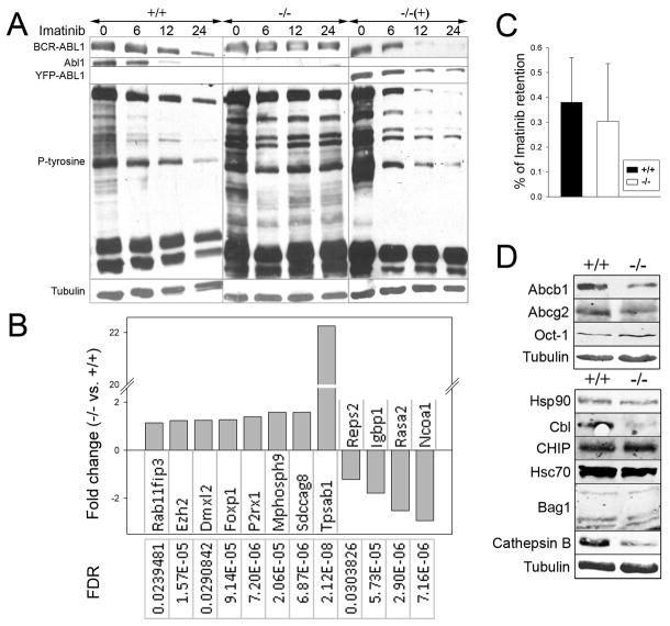
Figure 4. Imatinib-resistant phenotype of BCR-ABL1 –positive Abl1−/− leukemia cells.
BCR-ABL1 –positive Abl1−/− leukemia cells (−/−), BCR-ABL1 –positive Abl1+/+ leukemia cells (+/+) and BCR-ABL1 –positive Abl1−/− leukemia cells reconstituted with YFP-ABL1 (−/− (+)) were used. (A) Western analysis of the total cell lysates from cells incubated with 1μM imatinib for 0, 6, 12 and 24 hrs. (B) Statistically significant (FDR<0.05) fold-changes (>1) of the expression of indicated genes in BCR-ABL1-positive Abl1−/− versus BCR-ABL1-positive Abl1+/+ samples. (C) Intracellular retention of imatinib; results represent mean percentages ± s.d. of total C14-imatinib. (D) Western blots of total cell lysates to detect imatinib transporters (upper box) and proteins involved in BCR-ABL1 degradation (lower box).

