Fig. 3.
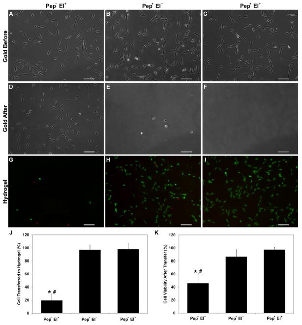
Cell transfer from gold substrate to hydrogel. Cells were transferred from non-modified substrates with electrical potential (Pep− El+), modified substrates without electrical potential (Pep+ El−), and modified substrate with electrical potential (Pep+ El+). Representative phase contrast images of the same substrate before (A–C) and after (D–E) transfer show negligible number of cells still adherent on peptide coated gold after transfer as opposed to the high number of cells on non-modified substrates. The corresponding fluorescent images (GFP/EthD-1) of the hydrogels after transfer display few rounded cells for the Pep− El+ (G) and numerous viable spread HUVECs for both Pep+ El− (H) and Pep+ El+ (I). (J) Mean percentage of cell transferred to hydrogel confirms that peptide modification enables complete HUVEC transfer as compared to only 20% of cells for Pep− El+. (K) Mean cell viability after transfer similarly illustrates that both modified substrates promoted transfer with high cell viability as opposed to non-modified ones (less than 50% viable cells). Gold surface coating with oligopeptide SAM enables HUVEC transfer to GelMA hydrogels with high efficiency and viability independent of electrochemical SAM desorption. (Scale bars: 100 μm; error bars: ± SD; statistically significant difference from Pep+ El− # and Pep+ El+ *)

