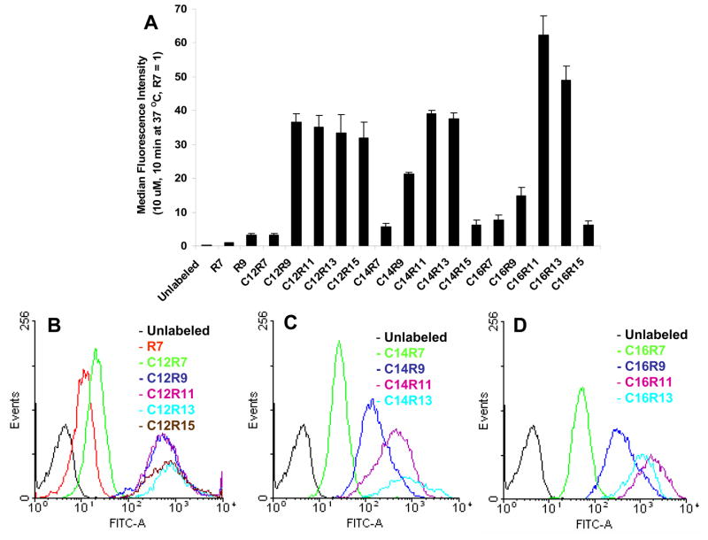
Fig. 1.
Cellular uptake of 10 μM LPs incubated with Jurkat cells for 10 min at 37 °C. (A) Jurkat cells incubated with LPs were washed, trypsin treated, PI stained, and analyzed by flow cytometry with 1–2 × 10⁴ events. (B, C, and D) The histograms of C12, C14, or C16 LPs. Each experiment was performed at least three times in triplicate, and the results are expressed as the median change in fluorescence ± S.D. The median fluorescence intensity of R7 in Jurkat cell was set as 1.

