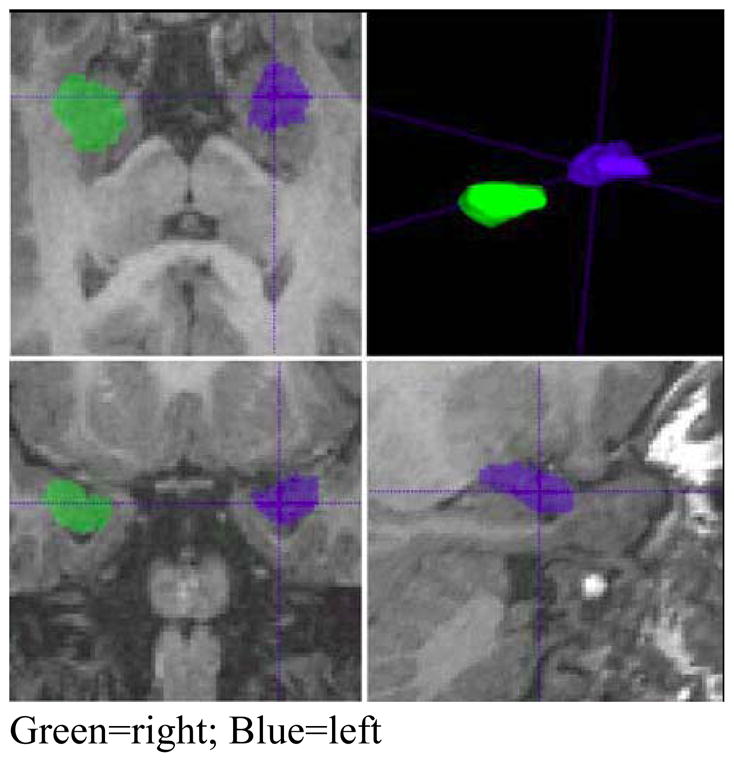
Figure 1.

Example of amygdala segmentations
Sample segmentation of right and left amygdala using IRIS software. Image is presented in radiological orientation with the right hemisphere visualized on the left side of the image (green) and the left hemisphere on the right side (blue).
