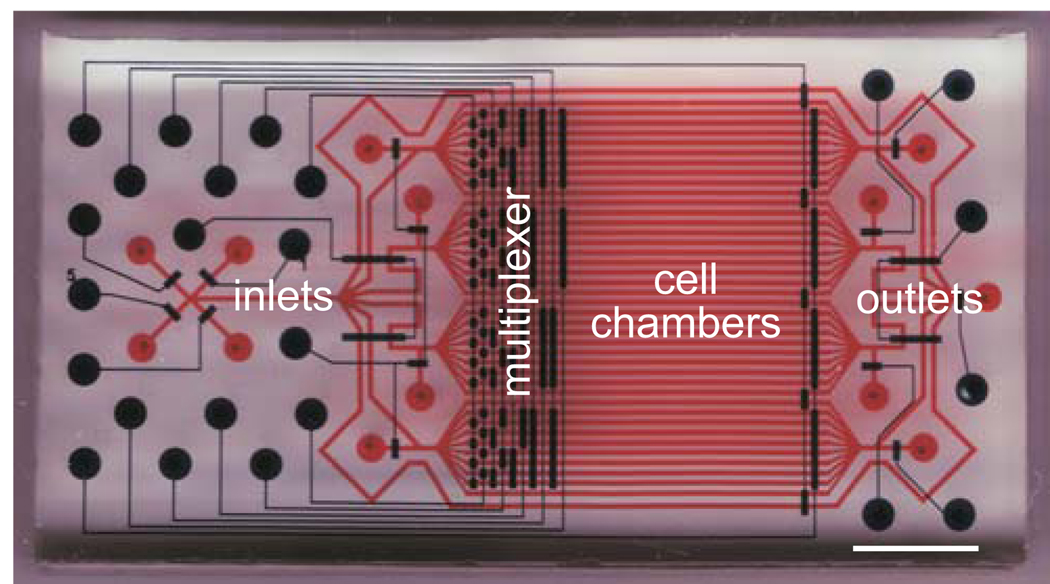
Figure 1. Design of a microfluidic device for high content screening.
Cell chambers and fluidic conduits are shown in red and integrated membrane valves are shown in blue. Device inlets, outlets, cell chamber, and multiplexed valves are indicated. Scale bar, 5 mm. This figure was originally published in Molecular Cellular Proteomics 19, and is used with permission.

