Figure 2.
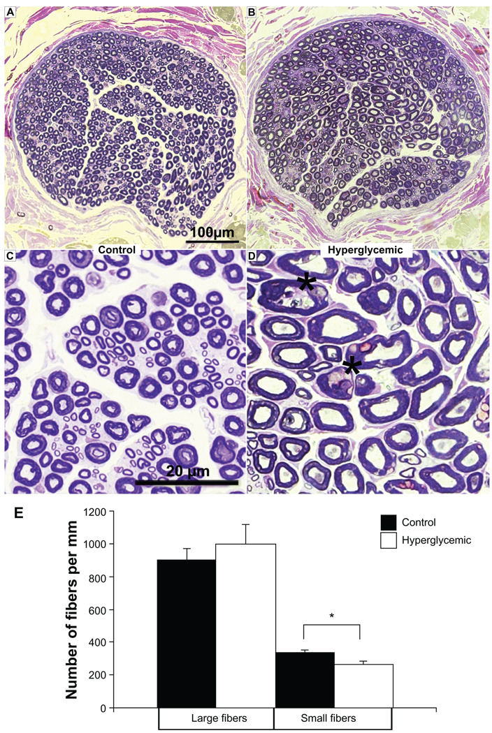
A, B) Sciatic nerve morphometry. Semi-thin toluidine blue sections prepared from control pig sciatic nerve reveal a dense network of large diameter myelinated fibers motor and small diameter myelinated sensory regularly-shaped fibers, whereas sections from a hyperglycemic sciatic nerve reveal visible loss of small fibers; overall fiber morphology is altered suggesting the onset of sensorimotor neuropathy. C, D) Lower panel images magnify nerve morphology, displaying large and small fibers in the control sections and indicating increase in the number of irregular shaped fibers (asterisks) in the diabetic nerve. E) Quantification of fiber number was performed on the indicated sections and reported as values per mm. *P < 0.05.

