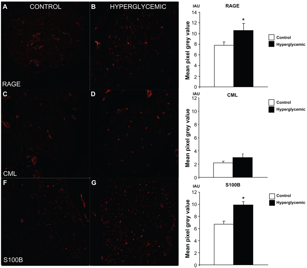
Figure 3.
Assessment of immunofluorescent signal in sciatic nerve sections single stained for RAGE and its ligands. Difference in immunofluorescent signal between control and hyperglycemic tissues was observed for RAGE (A, B) and S100B (E, F) indicating that under hyperglycemic conditions immunoexpression of these two proteins is increased. Level of CML (C, D) immunofluorescent signal was relatively low in both groups of animals and did not reach significant difference, however visible increase of CML positive fibers was noticed in hyperglycemic nerves suggesting that hyperglycemia likely induces immunoexpression changes of AGEs in the peripheral nerve over long period of time. *P < 0.05.

