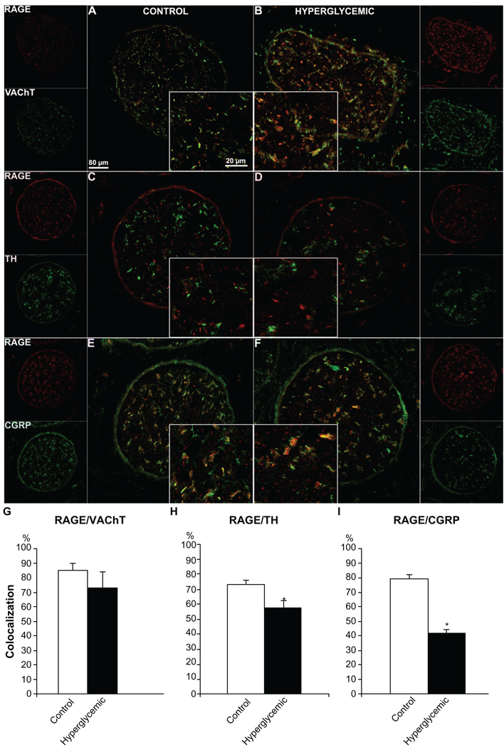
Figure 5.
Immunofluorescence analysis of RAGE distribution in different types of sciatic nerve fibers. Control and diabetic sciatic nerve tissues were subjected to RAGE (red) co-immunostaining with VAChT (motor neuron marker, A, B, green), TH (autonomic neuron marker, C, D, green) and CGRP (sensory neuron marker, E, F, green). Overlay images show that RAGE is localized in all types of sciatic nerve fibers. Note that the perineurial immunofluorescence was non-specific as observed in control staining with the secondary antibody alone. The quantitative analysis of the colocalization pattern revealed that although the number of double stained RAGE/VAChT positive fibers was similar in both groups of animals, there was a significantly lower number of RAGE/TH and RAGE/CGRP positive fibers between control and diabetic animals (G, H, I), supporting our earlier morphological findings on autonomic and sensory fiber loss in diabetic nerve. *P < 0.001.

