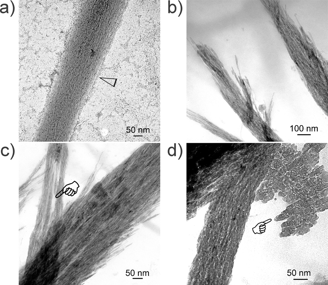
Figure 3.
Unstained TEM images showing different stages of non-periodic mineral assembly after reconstituted collagen was mineralized in simulated body fluid containing polyacrylic acid as a stabilization agent for amorphous calcium phosphate precursors. a) Initial mineralization at 24 h with calcium phosphate prenucleation clusters (open arrowhead) attaching to the fibril’s surface. b) Low magnification of the intrafibrillar deposits at 48 h. c) High magnification of the intrafibrillar deposits at 48 h showing strand-like intrafibrillar deposits. A region devoid of mineral deposition could be seen in one of the fibrils (pointer). d) Most of the fibrils at 72 h became heavily mineralized, with concomitant extrafibrillar mineralization observed in spaces between the mineralized fibrils. A less heavily mineralized fibril along the mineralization front is shown, showing non-periodic mineral deposition. Plate-like extrafibrillar crystals could be seen adjacent to the mineralized fibril (pointer).

