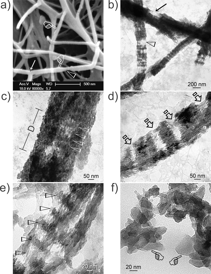
Figure 6.
a) SEM and b–f) unstained TEM of defective collagen-apatite nanocomposites when collagen is treated with TPP for 5 min. a) Defect along fibril surface (arrow) and a region with dehydration shrinkage in the absence of supporting minerals (arrowhead). b) A defect viewed from the side (arrow) and incomplete apatite deposition (open arrowhead). c) A defect (D) enables cross-banding to be seen within the subsurface (arrowheads) of a mineralized fibril. d) Regional mineralization of a fibril. Arrows: non-mineralized regions. e) Rotated fibril showing stacks of apatite platelets in the gap zones (arrowheads). f) Dissolution of the fibril’s organic phase with 6.7 mM NaOCl exposes the inorganic phase (pointers). Platelets are piled in a 3-D manner so that those at the back appear smaller than those in front.

