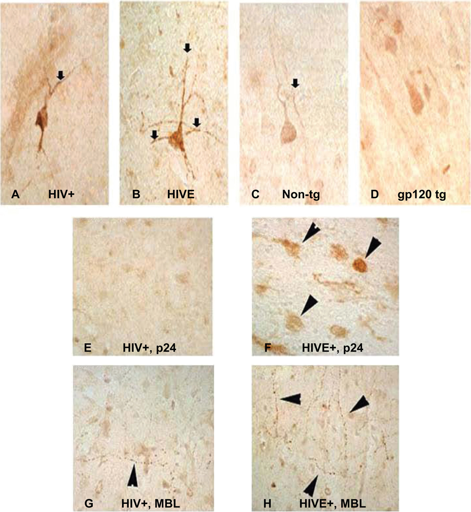
Figure 3.
Distribution of mannose binding lectin (MBL) in neuronal axons in human and mice brain tissues. Panels show MBL expression in single neuronal axon in postmortem brain tissues without (A) or with HIVE (B); while a comparative MBL expression in normal nontransgenic mouse (C) and gp120 transgenic mouse (D) is shown. Immunoreactive distribution of p24 in microglia of HIV+ non-HIVE (E) and HIVE case (F); and MBL in neuronal axons from HIV+, non-HIVE (G) versus HIVE (H) respectively are shown. Primary antibodies were developed with biotinylated secondary antibody, followed by treatment with Avidin D-HRP and reacted with 3, 3´-diaminobenzidine (DAB) in Tris buffer. Slides were counter-stained with 50% hematoxylene. 120 µm scale and 100× magnification was used.

